20x Signal Boost: New Brain Blood Flow Monitoring Breakth...
Scientists achieve a 20-fold signal boost in brain blood flow monitoring, enabling deeper tissue penetration and more accurate measurements for stroke care and neurosurgery.

Brain Health Depends on Blood Flow: How Can New Technology Detect Problems Faster?
Learn more about claude opus context generally available: what it means
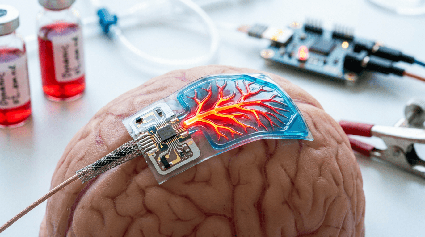
Brain health depends on steady blood flow delivering oxygen and nutrients to billions of neurons. When blood flow falters, brain function deteriorates within seconds. Scientists have now developed next-generation interferometric diffusing wave spectroscopy that achieves a 20-fold signal boost in cerebral blood flow monitoring, opening new possibilities for detecting and treating neurological diseases.
This breakthrough transforms how researchers and clinicians can observe the brain's vascular system in real time. The enhanced sensitivity allows for deeper tissue penetration and more accurate measurements, potentially revolutionizing stroke care, traumatic brain injury management, and neurosurgery.
What Is Interferometric Diffusing Wave Spectroscopy?
Interferometric diffusing wave spectroscopy represents an optical technique that measures blood flow through light behavior. When coherent light shines on living tissue, it creates speckle patterns that fluctuate as blood cells move through vessels. These "dancing" light grains reveal blood flow rates in the illuminated tissue volume.
The cerebral blood flow index (CBFi) quantifies this movement. Traditional methods struggled with weak signals and limited penetration depth, restricting their clinical utility. The next-generation approach solves these fundamental limitations through advanced interferometric detection.
How Does Light Reveal Blood Flow?
The physics behind this technology involves photon-tissue interactions. Coherent light photons scatter multiple times as they travel through brain tissue, encountering red blood cells, neurons, and other structures. Moving blood cells cause rapid fluctuations in the scattered light's phase and intensity.
Detectors capture these fluctuations and convert them into blood flow measurements. The faster the speckles fluctuate, the higher the blood flow rate. This non-invasive approach provides continuous monitoring without radiation exposure or contrast agents.
What Makes Next-Generation Technology 20 Times More Powerful?
For a deep dive on alicia keys at apple grand central: 50th anniversary event, see our full guide
The 20-fold signal enhancement stems from several technological innovations working in concert. Engineers optimized the interferometric detection scheme to capture more photons and reduce background noise. Advanced algorithms extract blood flow information from weaker signals that previous systems missed entirely.
Improved optical components increase light collection efficiency. The system uses specialized detectors with higher quantum efficiency and lower noise characteristics. These hardware improvements combine with sophisticated signal processing to achieve unprecedented sensitivity.
For a deep dive on disclosure day: spielberg's ufo film release & details, see our full guide
What Are the Key Technical Advantages?
The enhanced system delivers multiple benefits for cerebral blood flow monitoring:
- Deeper tissue penetration: Measures blood flow in brain regions previously inaccessible to optical methods
- Faster acquisition times: Captures rapid blood flow changes during neural activity or vascular events
- Higher spatial resolution: Pinpoints blood flow alterations in specific brain areas
- Reduced motion artifacts: Maintains accuracy even when patients move slightly
- Lower light exposure: Achieves better signals with less optical power, improving safety
How Can This Technology Help Stroke and Brain Injury Patients?
This technology addresses critical needs across multiple medical specialties. Neurologists can monitor stroke patients continuously, detecting secondary complications before irreversible damage occurs. Neurosurgeons gain real-time feedback during delicate procedures involving brain vasculature.
Intensive care units could deploy these systems for traumatic brain injury patients. Continuous cerebral blood flow monitoring helps clinicians optimize blood pressure management and detect dangerous intracranial pressure changes. Early intervention based on blood flow data may prevent secondary brain injury.
How Does It Improve Stroke Detection and Management?
Stroke represents a leading cause of death and disability worldwide. Rapid blood flow restoration determines patient outcomes. The enhanced monitoring system detects perfusion deficits immediately, guiding thrombolytic therapy decisions.
Clinicians can track reperfusion success in real time. If blood flow fails to improve after treatment, they can quickly pivot to alternative interventions. This immediate feedback loop shortens the critical window when brain tissue remains salvageable.
Why Do Neurosurgeons Need Real-Time Blood Flow Monitoring?
Brain surgery requires extreme precision to preserve healthy tissue while removing tumors or repairing aneurysms. Surgeons must avoid damaging blood vessels that supply critical brain regions. Real-time blood flow monitoring alerts surgical teams when their actions compromise perfusion.
The technology also assists in cerebrovascular bypass procedures. Surgeons can verify that new vessel connections provide adequate blood flow before completing the operation. This confirmation reduces postoperative complications and improves patient outcomes.
How Does the Signal Boost Improve Measurements?
The 20-fold signal enhancement translates directly into better clinical data. Weaker signals from deep brain structures become detectable and quantifiable. Researchers can now study blood flow in regions like the basal ganglia and thalamus, which play crucial roles in movement and cognition.
Increased signal strength also improves measurement reliability. Small blood flow changes that previously disappeared into background noise now stand out clearly. This sensitivity helps detect subtle perfusion problems before they cause symptoms.
How Deep Can the Technology Measure Blood Flow?
Traditional optical methods typically penetrate only 1-2 centimeters into brain tissue. The enhanced system reaches 3-4 centimeters or deeper, accessing a larger fraction of the cerebral cortex. This expanded coverage provides more comprehensive brain monitoring.
Accuracy improvements matter equally. The system distinguishes true blood flow changes from artifacts caused by scalp blood flow, skull anatomy variations, or environmental factors. Better signal quality means more trustworthy data for clinical decision-making.
What Can Researchers Learn About Brain Function?
Scientists studying brain function gain a powerful new tool for investigating neurovascular coupling. This phenomenon links neural activity to local blood flow increases, supplying active neurons with extra oxygen and glucose. Understanding this relationship helps explain how the brain maintains energy balance.
The technology enables studies of cerebral autoregulation, the brain's ability to maintain stable blood flow despite blood pressure fluctuations. Impaired autoregulation contributes to various neurological conditions. Better monitoring reveals when and why this protective mechanism fails.
How Will This Advance Neuroscience Understanding?
Researchers can now correlate blood flow patterns with cognitive tasks, sensory stimulation, or disease states. These investigations reveal how different brain regions communicate and coordinate their activities. The enhanced temporal resolution captures rapid blood flow dynamics during neural processing.
Longitudinal studies become more feasible with non-invasive, repeatable measurements. Scientists can track how aging, disease progression, or therapeutic interventions affect cerebral perfusion over months or years. This capability accelerates translational research bridging laboratory discoveries and clinical applications.
What Does Hospital Implementation Require?
Deploying this technology in clinical settings requires user-friendly designs and robust performance. The system must work reliably in busy hospitals with varying ambient light conditions and electrical interference. Engineers have prioritized portability and ease of use in the device architecture.
Cost considerations influence adoption rates. While the enhanced components increase manufacturing expenses, the non-invasive nature eliminates recurring costs for contrast agents or disposable sensors. Healthcare systems may find the technology cost-effective compared to alternatives like perfusion MRI or PET scanning.
How Much Training Do Clinical Teams Need?
Clinical staff need training to position optical sensors correctly and interpret blood flow data. However, the learning curve appears manageable compared to other neuroimaging modalities. Automated analysis algorithms reduce the expertise required for basic monitoring applications.
Integration with existing monitoring systems enhances utility. The cerebral blood flow data complements information from EEG, intracranial pressure monitors, and vital sign displays. Unified dashboards help clinicians synthesize multiple data streams for comprehensive patient assessment.
The Future of Brain Blood Flow Monitoring
Next-generation interferometric diffusing wave spectroscopy marks a significant advance in cerebral blood flow monitoring. The 20-fold signal boost enables deeper tissue penetration, faster measurements, and more accurate blood flow quantification. These improvements expand clinical applications from stroke care to neurosurgery to intensive care management.
Continue learning: Next, explore hammerspoon: automate your mac like a power user
The technology promises to improve patient outcomes by detecting perfusion problems earlier and guiding interventions more precisely. Researchers gain unprecedented ability to study brain function and neurovascular relationships. As the system transitions from laboratory to bedside, it may become an essential tool for protecting and preserving brain health across diverse medical scenarios.
Related Articles

iPhone 17 Unveiled: Features & Expectations
With the iPhone 17 launch just a week away, explore the rumored features, technological advancements, and Apple's push for sustainability.
Sep 5, 2025

Atlassian's Strategic Move: Acquiring The Browser Company
Atlassian's acquisition of The Browser Company marks a significant shift towards integrated, innovative digital workspaces and team collaboration tools.
Sep 4, 2025

Unpacking The Bitter Lesson in AI Evolution
Delving into the Bitter Lesson in AI: Unraveling its true meaning and impact on future technological innovations and trends.
Sep 4, 2025
Comments
Loading comments...